

An article on emedicine at http://www.emedicine.com/ped/topic1773.htm describes in PVL in more detail:
Quote
"Periventricular Leukomalacia
Article Last Updated: Feb 14, 2008
References Author: Terence Zach, MD, Department Vice-Chair, Professor, Department of Pediatrics, Section of Newborn Medicine, Creighton UniversityTerence Zach is a member of the following medical societies: American Academy of Pediatrics, American Medical Association, and Nebraska Medical AssociationCoauthor(s): James C Brown, MD, Codirector of Pediatric Radiology, Assistant Professor, Department of Radiology, Creighton University School of MedicineEditors: Scott S MacGilvray, MD, Associate Professor, Department of Pediatrics, East Carolina University School of Medicine; Mary L Windle, PharmD, Adjunct Assistant Professor, University of Nebraska Medical Center College of Pharmacy, Pharmacy Editor, eMedicine.com, Inc; Arun K Pramanik, MD, MBBS, Professor of Pediatrics, Director of Neonatal Fellowship, Louisiana State University Health Sciences Center; Carol L Wagner, MD, Professor of Pediatrics, Medical University of South Carolina; Ted Rosenkrantz, MD, Head, Division of Neonatal-Perinatal Medicine, Professor, Departments of Pediatrics and Obstetrics/Gynecology, University of Connecticut School of Medicine
Synonyms and related keywords: periventricular leukomalacia, PVL, ischemic brain injury, cerebral palsy, CP, hypotension, ischemia, coagulation necrosis, intracranial hemorrhage, ICH, hypocarbia, vasculitis, chorioamnionitis, cytokines, white matter damage, spastic diplegia, quadriplegia, nystagmus, strabismus, blindness, retinopathy of prematurity, maternal chorioamnionitis, respiratory distress syndrome, pneumonia, patent ductus arteriosus, placental vascular anastomoses, twin gestation, antepartum hemorrhage, sepsis
Background
Periventricular leukomalacia (PVL) is the most common ischemic brain injury in premature infants. The ischemia occurs in the border zone at the end of arterial vascular distributions. The ischemia of PVL occurs in the white matter adjacent to the lateral ventricles. The diagnostic hallmarks of PVL are periventricular echodensities or cysts detected by cranial ultrasonography. Diagnosing PVL is important because a significant percentage of surviving premature infants with PVL develop cerebral palsy (CP), intellectual impairment, or visual disturbances.
Pathophysiology
The pathophysiology of PVL is a complex process. PVL may occur because of ischemia-reperfusion injury to the periventricular area of the developing brain or because of cytokine-induced damage following maternal or fetal infection.
PVL is a white matter lesion in premature infants that may result from hypotension, ischemia, and coagulation necrosis at the border or watershed zones of deep penetrating arteries of the middle cerebral artery. Decreased blood flow affects the white matter at the superolateral borders of the lateral ventricles. The site of injury affects the descending corticospinal tracts, visual radiations, and acoustic radiations. In addition to possible ischemic injury, PVL may be the result of edema fluid and hemorrhage that cause compression of arterioles in the white matter. Reperfusion injury by free radicals to developing oligodendrocytes in the fetal or premature infant's brain may play an important role in the pathogenesis of PVL.
Premature infants have impaired cerebrovascular blood flow autoregulation and are susceptible to intracranial hemorrhage (ICH) as well as PVL. Premature infants on mechanical ventilation may develop hypocarbia. Several studies have linked hypocarbia, particularly in the first few days of life, with the development of PVL.1, 2
The relationship of maternal infection, placental inflammation, and vasculitis to the pathogenesis of PVL remains controversial. Some investigators have demonstrated an association of chorioamnionitis and cytokines with PVL although others have not.3
Following the initial insult, whether ischemia-mediated or cytokine-mediated, white matter damage occurs. The white matter damage likely occurs because of selective loss of oligodendrocytes.
Frequency
United States
Incidence of PVL ranges from 4-26% in premature infants in neonatal intensive care units (NICUs).
Incidence of PVL is much higher in reports from autopsy studies of premature infants.
As many as 75% of premature infants have evidence of PVL on postmortem examination.
Mortality/Morbidity
Cerebral palsy: Approximately 60-100% infants with PVL later develop signs of CP. Spastic diplegia is the most common form of CP following mild PVL. Severe PVL is frequently associated with quadriplegia.
Intellectual impairment: Varying degrees of intellectual impairment, developmental impairment, or both have been reported in association with PVL.
Visual dysfunction: Fixation difficulties, nystagmus, strabismus, and blindness have been associated with PVL. Some cases of visual dysfunction in association with PVL occur in the absence of retinopathy of prematurity, suggesting damage to optic radiations as causation.
Age
PVL occurs most commonly in premature infants younger than 32 weeks' gestation at birth.
CLINICAL
History
Periventricular leukomalacia (PVL) occurs most commonly in premature infants born at less than 32 weeks' gestation who have a birth weight of less than 1500 g. Many of these infants have a history of maternal chorioamnionitis. Most affected infants experience cardiorespiratory problems, such as respiratory distress syndrome or pneumonia, in association with hypotension or patent ductus arteriosus during their first days of life. Bacterial infection at birth also appears to be a risk factor.
Physical
Initially, most premature infants are asymptomatic. If symptoms occur, they usually are subtle. Symptoms may include the following:
Decreased tone in lower extremities
Increased tone in neck extensors
Apnea and bradycardia events
Irritability
Pseudobulbar palsy with poor feeding
Clinical seizures (may occur in 10-30% of infants)
Causes
Mechanically ventilated premature infants born at less than 32 weeks' gestation are at greatest risk for PVL.
Hypotension, hypoxemia, and acidosis may result in ischemic brain injury and PVL.
Marked hypocarbia in ventilated premature infants has been associated with increased risk of developing PVL.
Other associated risk factors include the following:
Placental vascular anastomoses, twin gestation, antepartum hemorrhage
Chorioamnionitis and funisitis
Sepsis
Maternal cocaine abuse
DIFFERENTIALS
Other Problems to be Considered
Intraventricular hemorrhagePeriventricular hemorrhagic venous infarction
Imaging Studies
Cranial ultrasonography
Cranial ultrasonography is the modality of choice for the initial evaluation of hypoxic-ischemic damage of the CNS in premature infants. Ultrasonography may be performed in the NICU without the need to transport fragile infants.
The earliest ultrasonographic appearance of periventricular leukomalacia (PVL) is abnormal increased echotexture in the periventricular white matter. This is a nonspecific finding that must be differentiated from the normal periventricular halo and mild periventricular edema that may not result in permanent injury.
The abnormal periventricular echotexture of PVL usually disappears at 2-3 weeks. Approximately 15% of infants experiencing PVL demonstrate periventricular cysts first appearing at 2-3 weeks after the initial increased echodensities.
The severity of PVL is related to the size and distribution of these cysts. Initial cranial ultrasonographic findings may be normal in patients who go on to develop clinical and delayed imaging findings of PVL.
CT scanning: CT scanning is not a first-line modality in evaluating these fragile premature infants in the first weeks of life. CT scanning may be helpful to better evaluate the extent and severity of PVL. Findings include ventriculomegaly involving the lateral ventricles with irregular margins of the ventricles and loss of deep white matter.
MRI: Like CT scanning, MRI does not play a major role in the early evaluation of PVL. MRI is most helpful in monitoring infants with suspected PVL and evaluating infants who develop clinical signs suggestive of PVL. MRI demonstrates the loss of white matter, abnormal signal intensity of the deep white matter, and ventriculomegaly. MRI demonstrates thinning of the posterior body and splenium of the corpus callosum in severe cases of PVL. Volumetric MRI scanning is also helpful in determining the extent of injury to the descending corticospinal tracts. A relationship between the degree of injury to the descending corticospinal tracts as assessed by MRI and the severity of diplegia has been reported.
Other Tests
EEG
Histologic Findings
PVL lesions demonstrate widespread loss of oligodendrocytes and an increase in astrocytes.
TREATMENT
Medical Care
No medical treatment is currently available. Free radical scavengers are being investigated to determine if they have a role in preventing oligodendrocyte injury in periventricular leukomalacia (PVL).
Consultations
Infants with PVL require close neurodevelopmental follow-up after discharge from the hospital. Potential consultants include pediatricians, developmental specialists, neurologists, and occupational and physical therapists.
FOLLOW-UP
Further Outpatient Care
Developmental follow-up: Premature infants with evidence of periventricular leukomalacia (PVL) require close developmental follow-up because of the high association with CP.
Early intervention strategies carried out by occupational therapists or physical therapists may decrease symptoms and may increase the infant's motor function.
Deterrence/Prevention
Prevention of premature birth is the most important means of preventing PVL.
Prior to birth, diagnosing and managing chorioamnionitis may prevent PVL. In 1999, Baud et al reported that betamethasone administered to mothers at 24-31 weeks' gestation, before delivery, significantly reduced the risk of PVL, suggesting the possible effect of steroids on fetal inflammatory response.4, 5
Avoiding maternal cocaine abuse and avoiding maternal-fetal blood flow alterations has been suggested to minimize PVL.
Following delivery of a premature infant, attempts to minimize blood pressure (BP) swings and hypotension may also be beneficial in preventing PVL.
Avoidance of prolonged hypocarbia in the mechanically ventilated premature infant may be useful in the prevention of PVL.
Prognosis
Infants with PVL are at risk for development of neurodevelopmental deficits. Mild PVL is often associated with spastic diplegia. Severe PVL is associated with quadriplegia. Severe PVL is also associated with a higher incidence of intelligence deficiencies and visual disturbances.
MISCELLANEOUS
Medical/Legal Pitfalls
Timing of initial cranial ultrasonography can be useful in determining the timing of the insult. Cystic PVL has been identified on cranial ultrasounds on the first day of life, indicating that the event was prenatal rather than perinatal or postnatal.
MULTIMEDIA
Media file 1: Cranial ultrasound, coronal view, in 1-week-old premature infant. The periventricular echotexture is abnormally increased (greater than or equal to that of the choroid plexus), which is consistent with the early changes of periventricular leukomalacia (PVL). Courtesy of Matthew Omojola, MD.
Media type: Ultrasound
Media file 2: Cranial ultrasound, coronal view, in 1-week-old premature infant without periventricular leukomalacia (PVL). The periventricular echotexture is normal. Compare with Media file 1. Courtesy of Matthew Omojola, MD.
Media type: Ultrasound
Media file 3: Cranial ultrasound, coronal view, in a 3-week-old premature infant. Multiple bilateral periventricular cysts are typical of this stage of periventricular leukomalacia (PVL). Courtesy of Matthew Omojola, MD.
Media type: Ultrasound
Media file 4: Cranial ultrasound, sagittal view, in 3-week-old premature infant. Multiple periventricular cysts are typical of this stage of periventricular leukomalacia (PVL). Courtesy of Matthew Omojola, MD.
Media type: Ultrasound
Media file 5: Cranial CT scan, axial image, in a 5-week-old premature infant with periventricular leukomalacia (PVL). The ventricular margins are irregular, which is consistent with incorporation of the periventricular cysts of PVL. Mild ventriculomegaly and loss of the periventricular white matter is observed. Courtesy of Matthew Omojola, MD.
Media type: CT
Media file 6: Cranial CT scan, axial image, in 14-month-old with periventricular leukomalacia (PVL). Ventriculomegaly is limited to the lateral ventricles secondary to diffuse loss of periventricular white matter. Courtesy of Matthew Omojola, MD.
Media type: CT
Media file 7: Cranial MRI, T1-weighted axial image, in an 18-month-old with periventricular leukomalacia (PVL). The lateral ventricles are enlarged without hydrocephalus. The periventricular white matter is diminished. Courtesy of Matthew Omojola, MD.
Media type: MRI
Media file 8: Cranial MRI, T2-weighted axial image, in an 18-month-old with periventricular leukomalacia (PVL). Again, enlarged ventricles and loss of white matter are demonstrated. Also noted is the abnormal increased signal in the periventricular regions on this T2-weighted image. Courtesy of Matthew Omojola, MD.
Media type: MRI
Media file 9: Cranial MRI, sagittal T1-weighted image in the midline, in an 18-month-old with periventricular leukomalacia (PVL). Hypoplasia of the corpus callosum is present and is most evident, involving the body. Courtesy of Matthew Omojola, MD.
Media type: MRI
References
Okumura A, Hayakawa F, Kato T, et al. Hypocarbia in preterm infants with periventricular leukomalacia: the relation between hypocarbia and mechanical ventilation. Pediatrics. Mar 2001;107(3):469-75. [Medline]. [Full Text].
Wiswell TE, Graziani LJ, Kornhauser MS, et al. Effects of hypocarbia on the development of cystic periventricular leukomalacia in premature infants treated with high-frequency jet ventilation. Pediatrics. Nov 1996;98(5):918-24. [Medline].
Kaukola T, Herva R, Perhomaa M, et al. Chorioamnionitis and cord serum proinflammatory cytokines: lack of association with brain damage and neurologic outcomes in very preterm infants. Pediatr Res. 2005;[Medline].
Baud O, Foix-L'Helias L, Kaminski M, et al. Antenatal glucocorticoid treatment and cystic periventricular leukomalacia in very premature infants. N Engl J Med. Oct 14 1999;341(16):1190-6. [Medline].
Canterino JC, Verma U, Visintainer PF, et al. Antenatal steroids and neonatal periventricular leukomalacia. Obstet Gynecol. Jan 2001;97(1):135-9. [Medline].
Bass WT, Jones MA, White LE, et al. Ultrasonographic differential diagnosis and neurodevelopmental outcome of cerebral white matter lesions in premature infants. J Perinatol. Jul-Aug 1999;19(5):330-6. [Medline].
Baud O, d'Allest AM, Lacaze-Masmonteil T, et al. The early diagnosis of periventricular leukomalacia in premature infants with positive rolandic sharp waves on serial electroencephalography. J Pediatr. May 1998;132(5):813-7. [Medline].
Dammann O, Hagberg H, Leviton A. Is periventricular leukomalacia an axonopathy as well as an oligopathy?. Pediatr Res. Apr 2001;49(4):453-7. [Medline]. [Full Text].
Dammann O, Leviton A. Brain damage in preterm newborns: might enhancement of developmentally regulated endogenous protection open a door for prevention?. Pediatrics. Sep 1999;104(3 Pt 1):541-50. [Medline]. [Full Text].
de Vries LS, Regev R, Dubowitz LM, et al. Perinatal risk factors for the development of extensive cystic leukomalacia. Am J Dis Child. Jul 1988;142(7):732-5. [Medline].
De Vries LS, Van Haastert IL, Rademaker KJ, et al. Ultrasound abnormalities preceding cerebral palsy in high-risk preterm infants. J Pediatr. Jun 2004;144(6):815-20. [Medline].
Enzmann DR. Imaging of neonatal hypoxic-ischemic cerebral damage. In: Stevenson DK, Sunshine P, eds. Fetal and Neonatal Brain Injury: Mechanisms, Management, and the Risk of Practice. 2nd ed. Oxford, England: Oxford University Press; 1997:302-55.
Hahn JS, Novotony EJ Jr. Hypoxic-ischemic encephalopathy. In: Stevenson DK, Sunshine P, eds. Fetal and Neonatal Brain Injury: Mechanisms, Management, and the Risk of Practice. 2nd ed. Oxford, England:. Oxford University Press;1997:277-286.
Hayakawa F, Okumura A, Kato T, et al. Determination of timing of brain injury in preterm infants with periventricular leukomalacia with serial neonatal electroencephalography. Pediatrics. Nov 1999;104(5 Pt 1):1077-81. [Medline]. [Full Text].
Haynes RL, Baud O, Li J, et al. Oxidative and nitrative injury in periventricular leukomalacia: a review. Brain Pathol. 2005;15:225-233. [Medline].
Kuban K, Sanocka U, Leviton A, et al. White matter disorders of prematurity: association with intraventricular hemorrhage and ventriculomegaly. The Developmental Epidemiology Network. J Pediatr. May 1999;134(5):539-46. [Medline].
Leviton A, Paneth N, Reuss ML, et al. Maternal infection, fetal inflammatory response, and brain damage in very low birth weight infants. Developmental Epidemiology Network Investigators. Pediatr Res. Nov 1999;46(5):566-75. [Medline].
Liao SL, Lai SH, Chou YH, Kuo CY. Effect of hypocapnia in the first three days of life on the subsequent development of periventricular leukomalacia in premature infants. Acta Paediatr Taiwan. Mar-Apr 2001;42(2):90-3. [Medline].
Murata Y, Itakura A, Matsuzawa K, et al. Possible antenatal and perinatal related factors in development of cystic periventricular leukomalacia. Brain Dev. 2005;27:17-21. [Medline].
Paul DA, Pearlman SA, Finkelstein MS, Stefano JL. Cranial sonography in very-low-birth-weight infants: do all infants need to be screened?. Clin Pediatr (Phila). Sep 1999;38(9):503-9. [Medline].
Shankaran S. Hemorrhagic lesions of the central nervous system. In: Stevenson DK, Sunshine P, eds. Fetal and Neonatal Brain Injury: Mechanisms, Management, and the Risk of Practice. 2nd ed. Oxford, England: Oxford University Press; 1997:151-64.
Volpe JJ. Brain injury in the premature infant: overview of clinical aspects, neuropathology, and pathogenesis. Semin Pediatr Neurol. Sep 1998;5(3):135-51. [Medline]. "
Unquote
This is our PRAYER, our HOPE, and our DREAM - to be a resource for special needs and adopted children and their families, especially those with unique and complex challenges, to make a difference in the life of a child, and in our journey to glorify JESUS. Every child has a story, and Every child matters.
"Defend the cause of the weak and fatherless; maintain the rights of the poor and oppressed."
Psalm 82 v 3
Please Donate and Help A Child
Consider helping a child with your donation?
By donating to a child, you will help to pay for that child's medical care, surgery, medication, doctor's visit, or medical equipment, school supplies, clothing, food and toys for their development?
All the children in listed here live within the United States. The families of these children have extra challenges in providing care for a variety of reasons such as unemployment, loss of medical insurance, life changing illness, life challenge or disability, extensive medical bills, or adoption.
Caring for five special children, we understand these daily challenges.
One time donations are acceptable through the donation button on our blog to the right at the top of our posts.
Every dollar goes to support both the child and administrative costs to continue our program.
One time donations in any amount are accepted ($5.00, $10.00, $20.00, $50.00, $100.00, $250.00, $500.00).
Weekly Donations are $10.00 !
Monthly Donations are $20.00 !
Donating is easy! We have various children right here in the United States who need your help. To donate to a child:
1. Click on the Donation button to the right at the top.
2. Choose how much you want to donate.
3. Write the child's name on the donation in comments.
4. Or Click on that Child's Donation button.
5. Your account will automatically be billed.
6. Subscribe to our blog for updates on how your child is doing.
7. Come back and donate when you want.
8. If you want to donate for a specific item, please write the child's name and the item in comments when you make the donation.
If you know of a child you recommend for sponsorship, please email us at academyofthepossible@gmail.com for more information. Be sure to include your email or mailing address, so we can send you the information.
Take a look at the children below. Each child has unique needs. Each child's needs will be updated as they change!
Michael: Michael is a charming young man who was adopted from Kazakhstan and suffered malnutrition in his early life due to the limited resources of the orphanage who did their best to care for him. Michael has multiple disabilities including Ectodermal Dysplasia, Chromose 16 deletion, Dysphagia, Cerebral Palsy, Juvenile Osteoporosis, Juvenile Rheumatoid Arthritis, Auditory Processing Disorder, Short Stature, Growth Hormone Deficiency, Cognitive and Developmental Delays, Speech and Language Delays, Thrombocytopenia, Hemolytic Anemia, Evan's Syndrome, Systemic Lupus, Hashimoto's Thyroiditis, Kidney Disease, Seizures, Autism, Periventricular Leukomalacia, Hypoplasia of the Corpus Callosum, Heart Murmur, Asthma, GERD, Gastrointestinal Disorder, medication, and on-going follow up, physical therapy, occupational therapy, medical equipment such as wheelchair, AFO braces, walker, cooling vest. Michael cannot play contact sports, so access to swimming for low impact exercise is ideal. A wheelchair accessible van would also help greatly.
Michael's Needs: Michael needs a phase cooling hat to help him keep cooler during the summer months and warmer weather, and his house needs new concrete for handicap accessible entrance to his home. Michael would also like a saltwater pool so he can swim at home in the evening when he won't be affected by the sun or heat or chlorine which can all cause him seizures. A new helmet for seizures. This will allow Michael to go outside more often. Michael would like a laptop and software to help him with his schoolwork, particularly Rosetta Stone Latin for High School, Teaching Textbooks Math, and All the Apologia Science on CD.
Michael's Dreams: Michael dreams of owning a guitar and becoming a medical doctor to help other kids like himself.
Gabriela: Gabriela is a sensitive young lady who was adopted from the Ukraine, given up for adoption again by her first family and then adopted by her forever family. Gabriela has Bipolar Disorder, Headaches, Frequent Episodes of Blindness, Muscle Twitches, Difficulty with Numbness, Tingling and Walking, Heartburn, Swallowing, Breathing, and also has ADHD, medication, on-going follow up. Gabriela is currently being evaluated for Multiple Sclerosis.
Gabriela's Needs: Gabriela needs regular donations to help pay for her physician follow up which insurance does not pay for. Gabriela also needs AFO braces to help her walk better.
Gabriela's Dreams: Gabriela dreams of rescuing animals and become a Veterinarian. Gabriela would really like some new art supplies and a new DS with all the pet games.
Joseph: Joseph was adopted from Estonia by his first family who was unable to care for him due to extenuating circumstances and then adopted by his forever family. Joseph is a charming young man who loves attention and has Severe Global Delays, FAS (Fetal Alcohol Syndrome), Pulmonary Hypertension, Transposition of the Major Vessels Repaired, several open heart surgeries to repair his declining heart over the next few years and a possible pacemaker, Cognitive Delays, Speech and Language Delays, Developmental Delays, Learning Disability, Microcephaly, Craniostenosis, Short Stature, Growth Hormone Deficiency, GERD, medication, and on-going follow up.
Joseph's Needs: Joseph is slow to grow and wears out his clothes and needs new shoes. An ipad that can help him develop more fully academically.
Joseph's Dreams: Joseph loves cars and dreams of working with the police when he grows up.
Joshua: Joshua also was adopted from Estonia and given up by his first family and later adopted by his forever family. Joshua is a quiet young man with Probable Fetal Alcohol Effects, Rough Beginnings, and Development and Learning Delays.
Joshua's Dreams: Joshua dreams of building rockets, piloting a plane and becoming an engineer.
Shawna: Shawna is a lovely girl adopted in the US who is healthy but lives with several siblings with special needs.
Shawna's Needs: Shawna needs new clothes, and shoes.
Shawna's Dreams: Shawna dreams of having her own horse to name Flicka, a pool to swim in, having new princess shoes and a princess dress, and lots of fruit trees to eat from in her backyard.
THANK YOU FOR YOUR KINDNESS, LOVE AND GENEROSITY!
Our Business
Our business Richardson Studios is a resource for parents particularly of special needs children. Come visit our webpage at http://www.richardsonstudios.com/
Our vision beginning the planning process is a school for special needs children. Read more at http://www.richardsonstudios.com/. Please consider a donation.
Our vision beginning the planning process is a school for special needs children. Read more at http://www.richardsonstudios.com/. Please consider a donation.
Our Webpage
Shop Online Richardson Studios
http://www.richardsonstudios.com
All money raised at our online store goes to support our vision of a special needs school. Check out our products. We have original paintings, used items, and much more coming. We are building our inventory some check back soon.
All money raised at our online store goes to support our vision of a special needs school. Check out our products. We have original paintings, used items, and much more coming. We are building our inventory some check back soon.
Visit Our Store ! Support Special Needs Kids
If there were a Catholic school for all special needs children from preschool to 12th with onsite services for various needs such as occupational or physical therapy, speech and language, psychology etc, would you or someone you know enroll a child?
Gifting
Towards a child in need, towards an adoption, and a child's dream of having a family. Money raised will help pay expenses such medical care and clothes. Please support our mission to help children around the world.
Thousands of children are dying daily from starvation and lack what we take for granted (the basics such as electricity, running water, heat, and education). Hundreds of thousands of children live in orphanages. Children are institutionalized because of special needs. Adoption is our mission field.
For many children, the simple desire is to be wanted and have a family of their own and to be loved.
Please consider a gift of $5.00, $10.00, or $20.00 and change the course of a child's life forever. Using the Donation button near the bottom of the blog posts is easy.
Small amounts are very welcome. Please consider even just $1.00. - which will go a long way to help a child.
Our Growing Family
"Defend the cause of the weak and fatherless; maintain the rights of the poor and oppressed." Psalm 82 v 3
We Adopted Five Kids !
Thank you for joining us on our journey !
May God's peace shine upon you, His everlasting love renew you, His grace uphold you, and His blessings pour out upon you.
The Richardson Family
May God's peace shine upon you, His everlasting love renew you, His grace uphold you, and His blessings pour out upon you.
The Richardson Family
Favorite Songs
- Be Strong and Courageous by Michael W Smith
- Desert Rose by White Heart
- The Man From Calvary by Jennifer Richardson
- I Will Be Here by Steven Curtis Chapman
- Breathe by Michael W Smith
- This is the Great Adventure by Steven Curtis Chapman
- Jesus Take The Wheel by Carrie Underwood
- Mr. Mom by Lone Star
Favorite Quotes
- "We Are Called To Be Adopted: He predestined us to be adopted as his sons through Jesus Christ, in accordance with his pleasure and will." Ephesians 1:5
- "Adoption has a way of emptying our pockets."
- "My goal and prayer in life is to be half as loving as Mother Theresa." Jennifer Richardson
- "Adoption is one of the best ways to lose that man-pleasing desire we carry that want(s) others to approve of us and what we do." Jenny
- "Sometimes I think it is not God saying no to us, but us giving up too soon when the fight got tough." Jennifer Richardson
- I will not leave you as orphans; I will come to you. John 14:18
- Delight yourself in the LORD and He will give you the desires of your heart. Commit your way to the LORD; trust in Him and He will do this: He will make your righteousness shine like the dawn, the justice of your cause like the noonday sun. Psalm 37:4-6 (NIV)
- God give us all the spirit of wisdom and revelation. Ephesians 1:14
- May the God of hope fill you with all joy and peace as you trust in him, so that you may overflow with hope by the power of the Holy Spirit. Romans 15:13
- For the vision is yet for the appointed time; it hastens toward the goal, and it will not fail. Though it tarries, wait for it; for it will certainly come, it will not delay. Habakkuk 2:3
- "We can make a difference - one child at a time." Jennifer Richardson
Prayers & Praises
PRAYER REQUESTS:
Safety and health for our little ones in Africa. - Ongoing
For The Lord's leading and ears to hear. - Ongoing
For The Letting Go God Asks of Us, - Ongoing.
Money and financial provision for the adoption. - Ongoing
For the homeless and hungry people of Africa. - Ongoing.
For those dying of starvation and malnutrition in Africa each moment. - Ongoing.
For the Lord to bless abundantly John and Laurie in their ministry. - Ongoing
For Mary to experience God's presence in every day, - Ongoing.
For all marriages and relationships everywhere, that God would be the center of the relationship, - Ongoing.
For everyone who suffers from Diabetes and Chronic conditions of any kind, that God would heal their bodies and bring them comfort, - Ongoing.
For everyone who suffers from any mental illness or psychiatric condition, depression, Bipolar Disorder, and ADHD, that the Lord would calm their minds and their souls and bring order, - Ongoing.
For the Chan's and the health of their children born and unborn, - Ongoing.
For the Szolomayer Family that the Lord may give them joy and for all their personal intentions - Ongoing.
For all families everywhere for peace and blessings during the Christmas season, that we may all grow closer to God's heart, - Ongoing.
For our family and God's protection, - Ongoing.
For our relatives to receive God's heart and truth, - Ongoing.
For the Lee Family that the Lord may give them joy and for all their personal intentions - Ongoing.
For all of our children for health, growth and peace, - Ongoing.
For Face to Face Ministries and God's leading and blessing, - Ongoing.
PRAISE REPORTS:
For Face to Face Ministries and all the staff who seek the Lord 12-2007
For our friends R and T who hope to finalize the adoption of this beautiful little boy. 12-2007
For my sister who traveled safely during the holidays.
For Scott who received healing from a cold. 11-2007
For Jennifer who received healing from her asthma 11-2007
For our Saturn which got a new tire 11-2007
For Shawna who received healing from her first cold at 20 months 11-02-07.
For Michael who got his cast off 11-13-07
Everyone got over their winter colds 11-14-07
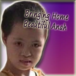
Anah
SCRIPTURES ON ADOPTION
Psalm 106:35 (Whole Chapter) but they mingled with the nations and adopted their customs.
Romans 8:23 (Whole Chapter) Not only so, but we ourselves, who have the firstfruits of the Spirit, groan inwardly as we wait eagerly for our adoption as sons, the redemption of our bodies.
Romans 9:4 (Whole Chapter) the people of Israel. Theirs is the adoption as sons; theirs the divine glory, the covenants, the receiving of the law, the temple worship and the promises.
Ephesians 1:5 (Whole Chapter) he [ Or sight in love. 5 He] predestined us to be adopted as his sons through Jesus Christ, in accordance with his pleasure and will—
SCRIPTURES ON GOD'S PROVISION
Romans 11:22 (Whole Chapter) Consider therefore the kindness and sternness of God: sternness to those who fell, but kindness to you, provided that you continue in his kindness.
Genesis 42:25 (Whole Chapter) Joseph gave orders to fill their bags with grain, to put each man's silver back in his sack, and to give them provisions for their journey. After this was done for them
Genesis 45:21 (Whole Chapter) So the sons of Israel did this. Joseph gave them carts, as Pharaoh had commanded, and he also gave them provisions for their journey.
Genesis 45:23 (Whole Chapter) And this is what he sent to his father: ten donkeys loaded with the best things of Egypt, and ten female donkeys loaded with grain and bread and other provisions for his journey.
Joshua 9:11 (Whole Chapter) And our elders and all those living in our country said to us, 'Take provisions for your journey; go and meet them and say to them, "We are your servants; make a treaty with us." '
Psalm 132:15 (Whole Chapter) I will bless her with abundant provisions; her poor will I satisfy with food.
Psalm 144:13 (Whole Chapter) Our barns will be filled with every kind of provision. Our sheep will increase by thousands, by tens of thousands in our fields
Proverbs 6:8 (Whole Chapter) yet it stores its provisions in summer and gathers its food at harvest.
Psalm 111:5 (Whole Chapter) He provides food for those who fear him; he remembers his covenant forever.
Proverbs 31:15 (Whole Chapter) She gets up while it is still dark; she provides food for her family and portions for her servant girls.
Luke 12:33 (Whole Chapter) Sell your possessions and give to the poor. Provide purses for yourselves that will not wear out, a treasure in heaven that will not be exhausted, where no thief comes near and no moth destroys.
Acts 11:29 (Whole Chapter) The disciples, each according to his ability, decided to provide help for the brothers living in Judea.
Acts 14:17 (Whole Chapter) Yet he has not left himself without testimony: He has shown kindness by giving you rain from heaven and crops in their seasons; he provides you with plenty of food and fills your hearts with joy."
Blog Archive
-
▼
2008
(216)
-
▼
June
(52)
- Update on Joseph's Heart Surgery !!!!!!!!!!!!!!!!!...
- Update on Michael
- Joshua
- Haircuts...
- HAIRCUT DAY !
- Special Moments -
- Birthday Pics...
- Birthday Pics...
- More Joseph Birthday Pics
- More Joseph Birthday Pics
- More of Joseph's Birthday Pics
- More Joseph's Birthday Pics
- More Joseph's Birthday Pics
- More of Joseph's Birthday Pics
- Joseph's Birthday Pics Are Here !!! June 11, 2008 ...
- Update on Joseph's Heart Surgery
- Waiting, waiting
- Update on Michael, Joseph, Joshua, Gabriela and Sh...
- An article on emedicine at http://www.emedicine.co...
- FAQ's on DCC
- DCC or Disorders of the Corpus Callosum
- NINDS Periventricular Leukomalacia Information
- Hypoplasia of the Corpus Callosum Information
- NINDS Craniostenosis Information
- NHANES Microcephaly
- Head Circumference
- Joseph's Genetics Appointment
- Joseph's Birthday Pictures Coming Soon !
- Upcoming Appointments
- Praises
- Michael's Cortisol Levels
- Joseph's Heart
- Praying for Joshua's Eye
- Update on Michael
- Gabriela
- Joshua
- Update on Shawna
- Update on Joseph
- The Flu: Yuck !!!!!!!!!!!!!!!!!!!!!
- Joseph (Tristan) is in the pool to the left. He co...
- Sunday Summer Fun: June 1, 2008
- NEW CLOTHES !!!
- May Birthday Celebrations...The Conclusion.
- May Birthdays...
- May Birthdays...
- May Brithdays continued...
- May celebrations...
- May Celebration...
- May Birthday Celebration continued...
- May Birthday Celebration continued...
- Celebrating May Birthdays -- May31, 2008
- Joshua in his fancy new shoes. Joseph in his fan...
-
▼
June
(52)
Site Counter
Email and Mailing List
If you would like to join our email list or mailing list, to receive updates and news on the Richardson Family, please send us an email to
academyofthepossible@gmail.com
with the words "Richardson Mailing List" in the subject line
with the following information:
Your first and last name
Your mailing address
Your city, state and zip code
Your email address
We look forward to hearing from you.
Blessings to you and your family,
The Richardson Family

Spiritual Resources
Shop.RichardsonStudios.com
Come check out our store !!! We have paintings, music, and used items.
http://shop.richardsonstudios.com/
Jennifer Richardson's Original Artwork is for Sale, and All items are originals and signed.
ALL PROCEEDS SUPPORT THE SPECIAL NEEDS CHILDREN.
Sizes vary from 4x6 to 11x14 and larger.
Paintings come in Watercolor, Acrylic, and Oil.
If you are interested, please email us at richardsonstudios@charter.net with the Word's "Jennifer's Artwork" in the subject line.
An Inspiration and Hope To All
" There is no such thing as doing Great Things in this World. We can only do small things with Great Love."
Mother Teresa
Mother Teresa
Adoption and Other Links
- Academy of the Possible Website
- Our Online Store - Richardson Studios
- Richardson Studios
- Academy of the Possible Support Group
- Adoption, General Information
- Bible
- Hall Family Blog
- A Child's Waiting
- Barbers
- Global Orphan Outreach
- Carlita's Website - mathonthelevel
- Ethiopia Facts
- Homeschool in the Little House Blog
- JCICS
- Jocelyn and Isaac
- Koinonia Academy Blog
- Liberia Map
- Liberia, Information On
- McKinney Blog
- National Geographic on Africa
- Shaohannah's Hope
- The Berry Patch Blog
- The Berry's Photo Blog
- World Partners Adoption
Facts on Africa
Africa is the second largest continent in the world made up of 53 independent countries. There is a great diversity in cultures and people. Africa is almost entirely surrounded by water and the land ranges from deserts to rain forests and mountains to grassland. National Geographic has a wonderful amount of information on their webpage about Africa. At the same time, at least 15 countries in Africa are the least developed in the world struggling with famine, disease, HIV and AIDs in some countries, extreme poverty, and lack of education. Maryknoll states that "70% of its population survives on less than $2 a day." I certainly could not live off of that.
BASIC FACTS
Continental Area - 11,700,000 square miles
Estimated Population - 690 million people
Largest City - Cairo, Egypt, 9.2 million people
Largest Country - Sudan, 968,000 square miles
Longest River - Nile, 4,160 miles
Largest Lake - Victoria, 26,828 square miles
Tallest Mountain - Kilimanjaro, Tanzania, 19,340 feet
MALARIA
According to the CDC, "In areas of Africa ..., an estimated 990,000 people died of malaria in 1995 – over 2700 deaths per day, or 2 deaths per minute."
AIDS EPIDEMIC
According to standwithafrica.org, "Africa is home to nearly 70% of adults and 80% of children living with HIV in the world, and has buried three-quarters of the more than 20 million worldwide who have died of AIDS since the epidemic began."
CHILD MARRIAGES - often driven by poverty:
According to the CDC, "In 2002, ≈52 million girls less than 18 years of age were married. With ≈25,000 girls less than 18 years being married each day, an estimated 100 million will be married by 2012... Child marriages occur 42% for Africa..."
THE MOST VULNERABLE
According to stand with africa, "In eastern Africa, Ethiopia, Kenya, and Somalia continue to suffer from prolonged periods of drought. The pastoral regions of these countries are the most vulnerable, and almost 2 million people in the Horn of Africa received emergency rations from World Food Program in 2001."
THE WORST DROUGHT
According to save the children, "Save the Children and other humanitarian organizations are calling the worst drought Africa has seen since 1984."
SUFFERING AFRICA
According to CNN, "Ethiopia ...Drought, AIDS and preventable disease have put millions of Africans at risk of starvation. People in southern Africa and the Horn of Africa stand to suffer most, officials say. "
RISK OF STARVATION
According to the UN, "World Food Program (WFP) said in December that more than 38 million people across Africa are at risk of starvation."
FOOD SHORTAGES
According to CNN, "Hardest hit are the Horn of Africa, where about 17.9 million people face severe food shortages, and southern Africa, where 16.41 million are at risk"
FOOD CRISIS
According to the UN, "In Ethiopia, ...expects to provide food aid to 11.3 million people in 2003, but that number could rise to 14.3 million people by year's end. "
A CONTINENT OF HUNGRY PEOPLE
According to Fr. Richard Roy, director of the Missionaries of Africa's Washington, DC, office, www.missionariesofafrica.org, "An entire continent of people are in dire need of food, clean water and affordable medicine. "
MASS STARVATION
James Morris, the Executive Director of the UN World Food Program used the words "mass starvation in Africa" and the UN referred to "Africa's critical situation".
CHILDREN STARVING TO DEATH
According to the BBC, "Children are dying daily in the few feeding centres there are, where their place in the queue could make the difference between life and death. " The BBC also said "Children are dying of starvation in feeding centres in Niger, where 3.6m people face severe food shortages"
TOGETHER WE CAN MAKE A DIFFERENCE. WE CAN DO SOMETHING. DONATE AND HELP US HELP THREE CHILDREN WHO NEED US.
Liberian Statistics
Liberia: means “land of the free”
Founded By: free African Americans and freed slaves from the USA in 1820
Civil War: 1989 to 2003, a total of 14 years
Below Poverty: more than 80%
Life Expectancy in the Total Population: only 39.65 years
Literacy in the Total Population: only 57.5%, and in the female population only 41.6%
Labor Population: is 70% agriculture
Unemployment: 85%
Population: > 3 million
Population Growth: 4.91%
Ethnics Groups Indigenous African tribes: 95% including Kpelle, Bassa, Gio, Kru, Grebo, Mano, Krahn, Gola, Gbandi, Loma, Kissi, Vai, Dei, Bella, Mandingo, and Mende
Ethnic Groups Americo-Liberians: 2.5% (descendants of immigrants from the US who had been slaves
Ethnic Groups Congo People: 2.5% (descendants of immigrants from the Caribbean who had been slaves)
Religious Beliefs: indigenous beliefs 40%, Christian 40%, Muslim 20%
Fertility Rate: 6.02 children per woman
Location: Western Africa, bordres the North Atlantic Ocean, between Cote d’lvoire and Sierra Leone
Total Square Miles Area: 111,370
Climate: tropical, hot, humid, dry winters with hot days and cool to cold nights; wet cloudy summers with frequent heavy showers.
Terrain: Mostly flat to rolling coastal plains rising to rolling plateau and low mountains in northeast
President: Ellen Johnson Sirleaf
Flag: Red and White striped with one white star on a blue background in the upper left corner (similar in style to the US flag)
Capital: Monrovia
United Nations: Member of
Currency: Primarily the Liberian Dollar (LD), also accepts US dollars
Major Languages: English, Ethnic group languages
Sources: Courtesy of the CIA World Factbook 2006 and the US Department of State Country Background Notes of 05-2007, and globaledge.msu.edu
No comments:
Post a Comment